![]() Figure 10 of
Mojumder, Mol Vis 2007;
13:2163-2182.
Figure 10 of
Mojumder, Mol Vis 2007;
13:2163-2182.
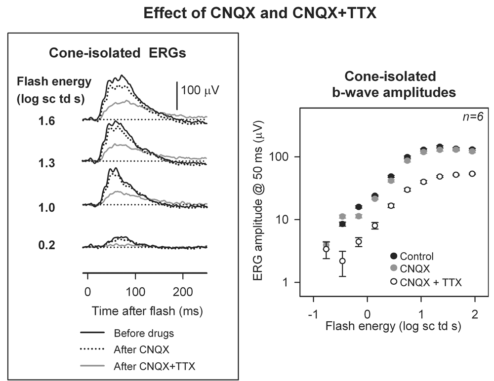
Figure 10. Effect of tetrodotoxin on the cone-driven electroretinogram arising from the cone to ON cone bipolar cell circuit
A: Dark-adapted isolated cone-driven responses are shown before intravitreal drug injections (black trace), after 6-cyano-7-nitroquinoxaline-2,3-dione (CNQX; dashed trace) and after tetrodotoxin (TTX) added following CNQX (CNQX+TTX; grey trace). B: Average electroretinogram (ERG) amplitudes are shown for all eyes for which the control condition, as well as both drug conditions were available (six eyes from four subjects). Amplitudes of ERGs were measured at 50 ms after the flash, the time to peak of the cone-isolated b-wave and plotted as a function of flash energy for the control dark-adapted cone-isolated b-wave (black circle), after CNQX (grey circle) and after CNQX+TTX (circle) plotted on a log-log scale.