![]() Figure 1 of
Mojumder, Mol Vis 2007;
13:2163-2182.
Figure 1 of
Mojumder, Mol Vis 2007;
13:2163-2182.
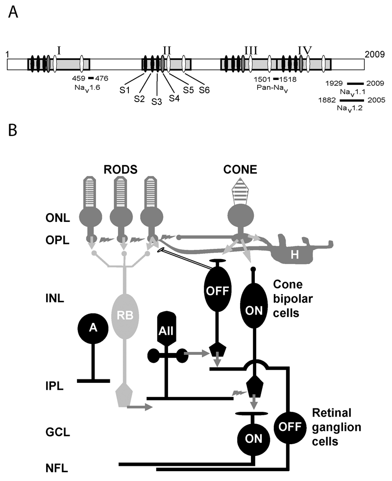
Figure 1. Voltage-gated Na+ channel (Nav) α-subunit structure and known distribution in the retina
A: Schematic representation of the voltage-gated Na+ channel (Nav) α-subunit. Light grey blocks (I-IV) indicate the highly conserved pore-forming regions within the α-subunit. Each contains six transmembrane domains (ovals, S1-S6). The fifth and sixth transmembrane domains (white ovals) line the channel pore and the fourth transmembrane domain (grey) is the voltage sensor. The amino acid residues targeted by the Pan-Nav α-subunit antibody and the subunit-specific amino acid sequences targeted by the Nav1.1, Nav1.2, and Nav1.6 antibodies used in this report are indicated along the bottom of the panel. After Yu and Caterall, 2003 [1]. B: Schematic diagram of Nav channel localization in rat retinal circuits based on physiological data. Black shading indicates cell types for which there is evidence for functional Nav channel expression. Functional Nav channels have been identified in subsets of ON and OFF cone bipolar cells, several subtypes of amacrine cells (including the AII subtype) and in ON and OFF retinal ganglion cells. Arrows indicate chemical synapses; lightning bolts indicate electrical synapses. The following abbreviations were used: outer nuclear layer (ONL); outer plexiform layer (OPL); inner nuclear layer (INL); inner plexiform layer (IPL); ganglion cell layer (GCL); nerve fiber layer (NFL); amacrine cell (A); horizontal cell (H); rod bipolar cell (RB).