![]() Figure 5 of
Morita, Mol Vis 2007;
13:2119-2128.
Figure 5 of
Morita, Mol Vis 2007;
13:2119-2128.
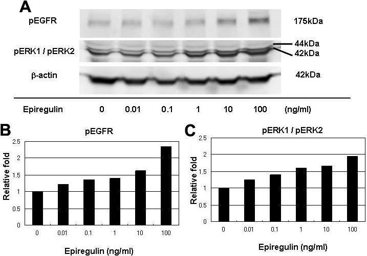
Figure 5. Phosphorylation of ErbB1 (EGFR) and ERK1/2 by epiregulin
HCECs were exposed to serial concentrations of epiregulin for 5 min. The cell lysates were subjected to 7.5% SDS-PAGE and then phosphorylated ErbB1 and ERK1/2 were detected with anti-phosphorylated-ErbB1 or anti-phosphorylated ERK1/2 antibody. The expression levels of ErbB1 and ERK1/2 were measured relative to that of β-actin in the same sample.