![]() Figure 2 of
Roger, Mol Vis 2007;
13:206-219.
Figure 2 of
Roger, Mol Vis 2007;
13:206-219.
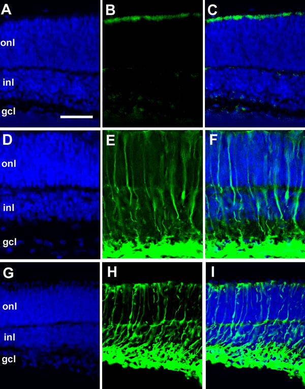
Figure 2. Glial fibrillary acidic protein induction in retinal explants after six days of cytokine treatment
P0 retinal explants were cultured with medium alone (control; A-C) or supplemented with 10 ng/mL of either ciliary neurotrophic factor (D-F) or leukemia inhibitory factor (G-I). After six days in vitro (6DIV) of culture, explants were fixed and stained with anti-glial fibrilary acidic protein (green). Gcl represents ganglion cell layer; inl represents inner nuclear layer; and onl stands for outer nuclear layer. Scale bar equals 40 μm.