![]() Figure 7 of
Okamoto, Mol Vis 2007;
13:2112-2118.
Figure 7 of
Okamoto, Mol Vis 2007;
13:2112-2118.
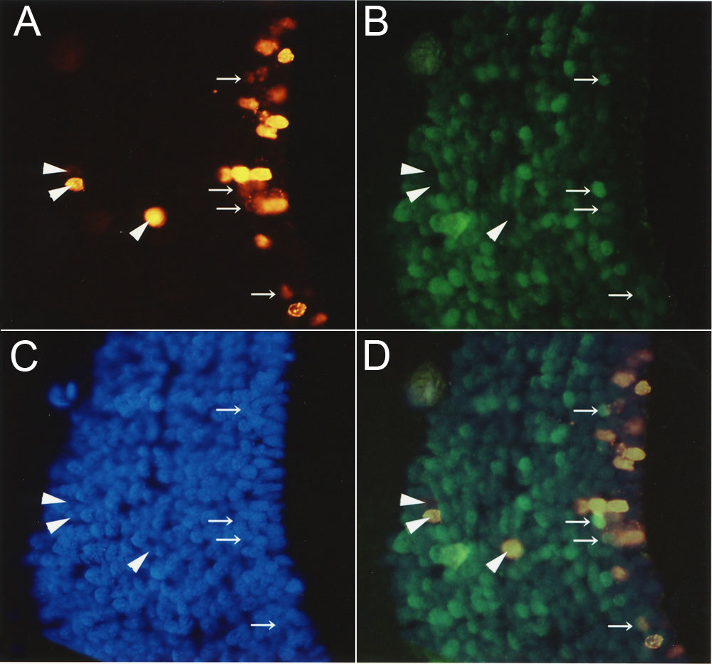
Figure 7. BrdU incorporation in cells of the regenerating brain two months after surgery
A: Cells staining positive for BrdU were mainly present in the ependymal cell layer. B, C: NeuN (neuronal marker) staining and DAPI staining, respectively. D: Shown is an overlay of A, B and C. Arrows point to NeuN-positive cells, and arrowheads mark NeuN-negative cells. NeuN-negative and BrdU-positive cells could be another cells type, such as astrocytes.