![]() Figure 5 of
Okamoto, Mol Vis 2007;
13:2112-2118.
Figure 5 of
Okamoto, Mol Vis 2007;
13:2112-2118.
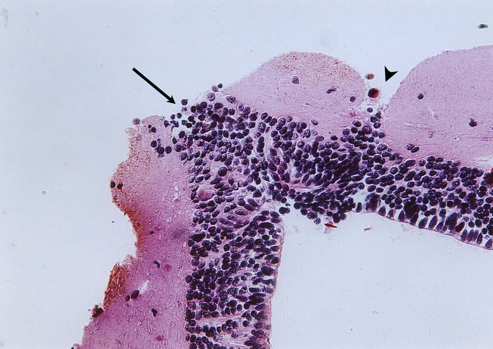
Figure 5. The coronal section of the optic tectum at one-month post surgery
This figure shows histological features of wound healing in the brain. This sample was taken one month after excision of the left tectum. The arrowhead indicates the joint of the left and the right optic tectum (OT), and the arrow indicates the wound closure. Wound closure of the lesion seemed to occur earlier than brain tissue regeneration.