![]() Figure 2 of
Okamoto, Mol Vis 2007;
13:2112-2118.
Figure 2 of
Okamoto, Mol Vis 2007;
13:2112-2118.
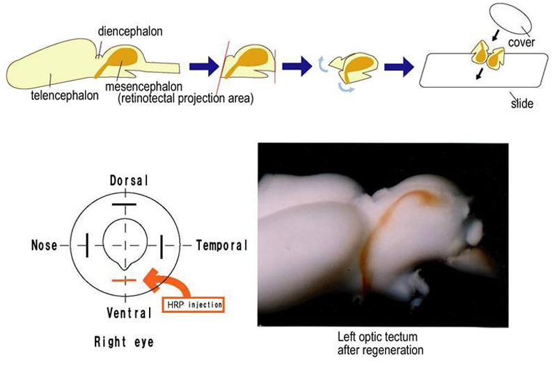
Figure 2. Method for detecting regeneration of the retinotectal projections
The top panel shows the method we devised for quantitation of regeneration of the retinotectal projections. The area that contains the regenerated tectum was excised from the brain. The brain was then divided in two parts by a slit in the ventral side. The right part was the uninjured part, while the left part was the injured and regenerating part. The flattened sides were placed on a glass slide and the photo prints were used to measure the area of the optic tectum that was innervated by the axons (see Methods). The lower panel on the left shows the injection of horseradish peroxidase (HRP) on the ventral site and on the right it shows how the HRP has been taken by the nerve fibers to indicate innervation in the regenerated optic tectum.