![]() Figure 5 of
Zhang, Mol Vis 2007;
13:2096-2104.
Figure 5 of
Zhang, Mol Vis 2007;
13:2096-2104.
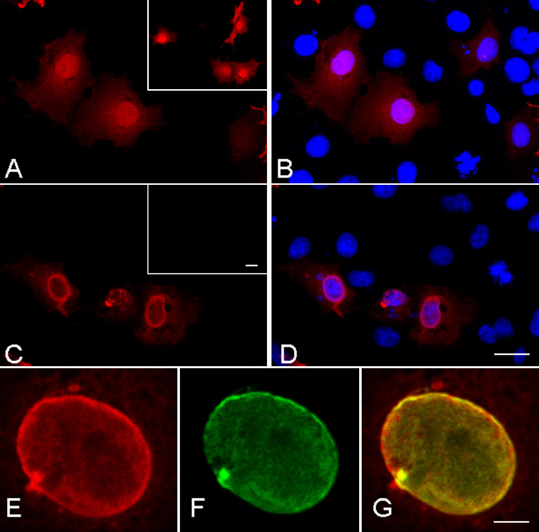
Figure 5. Localization of FLAG-tagged wildtype or G165fs γD-crystallin in COS-7 cells
A and B: Immunofluorescence of FLAG (red fluorescence) shows the distribution of wildtype γD-crystallin in both nucleus and cytoplasm. An overlay picture with DAPI-stained nucleus (blue fluorescence) is shown in B. Inset in A, similar distribution was detected by anti-γD-crystallin antibody. C and D: The G165fs mutant was localized in the nuclear periphery. Inset in C shows Negative staining was obtained by anti-γD-crystallin antibody which recognizes the COOH-terminal region. E-G: Confocal double immunofluorescence of FLAG (red fluorescence; E) and lamin A/C (green fluorescence; F) and their overlay (G) demonstrated colocalization of mutant γD-crystallin with lamins in the nuclear envelope. Scale bars: 20 μm (A-D) and 5 μm (E-G).