![]() Figure 7 of
Yuan, Mol Vis 2007;
13:2083-2095.
Figure 7 of
Yuan, Mol Vis 2007;
13:2083-2095.
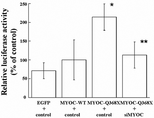
Figure 7. BiP activation as a stress response to myocilins in TM5 cells by luciferase reporter assays
BiPpGL3 vector was cotransfected in TM5 cells with either MYOCpEGFP (MYOC-WT) or MYOCQ368XpEGFP (MYOC-Q368X) along with control pH1-RNA or shMYOC-A. Dual luciferase assays were performed at 48 h after transfections. The results were from three independent transfection experiments and each experiment was tested in triplicates (n=9, bars=SD). The activity of BiP in TM5 cells after the cotransfection of MYOCpEGFP (MYOC-WT) and control pH1-RNA was used as a baseline control (100%, as seen in the second bar). Compared with the baseline, cotransfection of pEGFP (without MYOC) and the control, pH1-RNA, showed less activation of BiP in TM5 cells (as shown in the first bar), indicating that the presence of MYOC-WT could induce moderate stress response in TM5 cells. Significant activation of BiP was noted in TM5 cells after cotransfection of MYOCQ368XpEGFP (MYOC-Q368X) and control pH1-RNA plasmids, indicating that mutant MYOC-Q368X induced a more pronounced stress response than MYOC-WT (The asterisk indicates that p<0.001, as seen in the third bar). Cotransfection of shMYOC-A significantly reduced the activation of BiP induced by mutant MYOC-Q368X (The double asterisk indicates that p<0.001, as seen in the fourth bar), indicating shMYOC-A could reduce the stress response induced by the mutant myocilin to a baseline level similar to that induced by wild-type myocilin.