![]() Figure 5 of
Yuan, Mol Vis 2007;
13:2083-2095.
Figure 5 of
Yuan, Mol Vis 2007;
13:2083-2095.
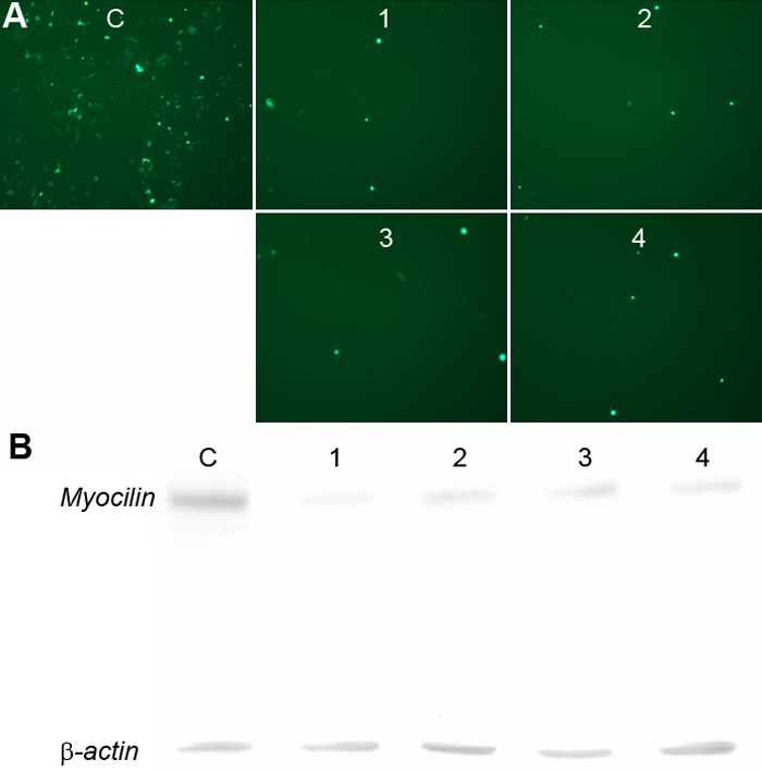
Figure 5. Suppression of MYOC-EGFP by short hairpin RNAs specific to various coding regions of MYOC in cultured HEK293 cells
A: Representative photographs of HEK293 cells from fluorescence microscopy after cotransfection of MYOCpEGFP with various shMYOCs are shown. The fluorescence photographs were taken with the same exposure times at 48 h after cotransfection. C: Cotransfection of MYOCpEGFP with a control plasmid; 1: Cotransfection of MYOCpEGFP with shMYOC-A; 2: Cotransfection of MYOCpEGFP with shMYOC-B; 3: Cotransfection of MYOCpEGFP with shMYOC-C; and 4: Cotransfection of MYOCpEGFP with shMYOC-D. Significant reduction of MYOC-EGFP was noted in HEK293 cells cotransfected with MYOCpEGFP and shMYOC-A, -B, -C, and -D, respectively (A 1-4). B: Western blot of protein lysates from cultured HEK293 cells after cotransfection of MYOCpEGFP with various shMYOCs as in A is shown. Equal amounts of protein lysates from HEK 293 cells (20 μg/lane) were loaded for each lane as indicated by similar intensities of β-actin in each lane. The MYOC-EGFP fusion protein and β-actin were detected with anti-EGFP and anti β-actin antibodies, respectively. Consistent with the findings of fluorescence microscopy in A, significant reduction of MYOC-EGFP was noted in HEK293 cells cotransfected with MYOCpEGFP and shMYOC-A, -B, -C, and -D, respectively (lanes 1-4). These protein bands were then digitized to quantify the suppression efficiency of each shRNA (see the Results section for average suppression efficiency of each shMYOC).