![]() Figure 5 of
Kottegoda, Mol Vis 2007;
13:2073-2082.
Figure 5 of
Kottegoda, Mol Vis 2007;
13:2073-2082.
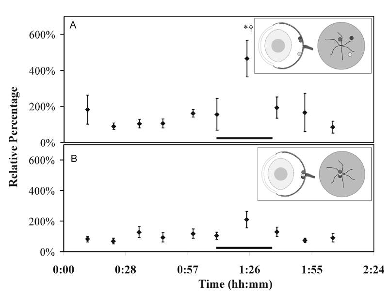
Figure 5. Different chemical response to different probe placement
Probe placement at VRI was confirmed by L-trans-pyrrolidine-2,4-dicarboxylic acid (PDC) infusion to specific regions of the retina. Plot of the relative levels of glutamate when the sampling probe tip was placed A 1-2 mm peripheral to the optic nerve head (n=3), or B directly over the optic nerve head (n=3). In the schematic drawings in the insets, dots mark the in vivo sampling sites and are shaded to show congruence between forward and cross section views. The increase in glutamate coincident with PDC infusion is significant at peripheral sampling sites both with respect to the basal levels, asterisk (*; p<0.05) and versus the glutamate increase seen at sampling sites over the optic nerve head, dagger (cross; p<0.05).