![]() Figure 2 of
Kottegoda, Mol Vis 2007;
13:2073-2082.
Figure 2 of
Kottegoda, Mol Vis 2007;
13:2073-2082.
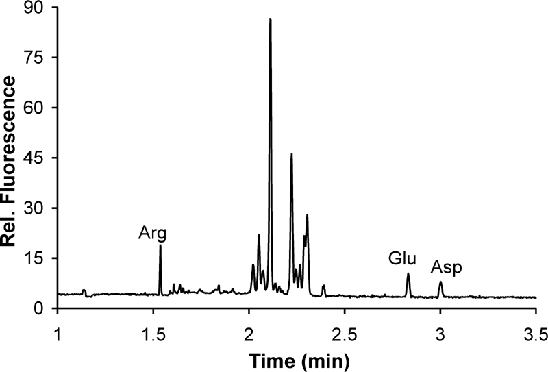
Figure 2. Fast electropherogram of rat vitreoretinal interface perfusate
Perfusate samples (500 nl) were mixed with 1 μl, 10 mM 3-(4-carboxybenzoyl) quinoline-2-carboxyladehyde (CBQCA) in dimethyl sulfoxide (DMSO) and then with 1 μl of 10 mM KCN. Electrophoretic separation analysis followed a 2 h incubation in the dark. Separation of Arg, Glu, and Asp was achieved in 3 min. The run buffer consisted of 20-mM sodium borate (pH 9.3), 45 mM SDS and 55 mM β-CD. A 41-cm long capillary was used with the detection window placed 30-cm from the inlet. The field strength during the separation was 490 V/cm.
