![]() Figure 1 of
Kottegoda, Mol Vis 2007;
13:2073-2082.
Figure 1 of
Kottegoda, Mol Vis 2007;
13:2073-2082.
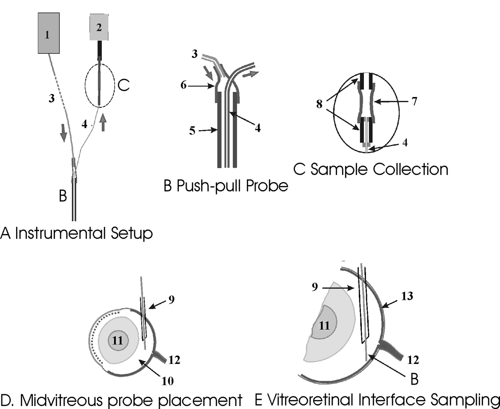
Figure 1. Details of basic instrumental set up and important features of sampling from rat eye
The entire push-pull perfusion system is shown in A. A programmable syringe pump (1) connected to the push-pull probe (B) via an infusion capillary (50 μm/150 μm inner diameter/outer diameter; 3) is that used to deliver Krebbs-Ringer buffer to the probe while perfusate is collected via a withdrawal capillary (20 μm/90 μm inner diameter/outer diameter; 4) through collection tubing (C) connected to a vacuum pump (2). The concentric design of the push-pull probe is shown in B. The infusion tubing (3) is connected to the larger outer probe capillary (110 μm/170 μm inner diameter/outer diameter; 5) through a segment of Tygon tubing (6). The inner withdrawal capillary in the probe (4) is connected to the vacuum pump through a sample collection tubing shown in C. The outlet of the withdrawing capillary and the inlet of the vacuum pump are connected by a 4-cm segment of Tygon tubing (7) that is used for collection and storage of perfusates. Stainless steel tubing (23-gauge) (8) is used to make a tight seal with the Tygon collection tubing. A schematic of the probe placement into the rat eye is shown in D. A 29-gauge guide needle (9) is inserted into the posterior chamber (10) at the extreme periphery of the retina. The eye is shown in cross section with the lens (11) and optic nerve head (12) labeled. The probe tip can also be positioned at the vitreoretinal interface shown in E. Under visual guidance the probe B is advanced to the retina (13) until barely in contact.