![]() Figure 1 of
Zamora, Mol Vis 2007;
13:2058-2065.
Figure 1 of
Zamora, Mol Vis 2007;
13:2058-2065.
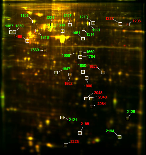
Figure 1. Representative overlaid gel of Cy dye-labeled retinal and choroidal endothelial protein samples
Differentially abundant protein spots that were visible in at least four of five donor gels were detected by the significance analysis of microarrays method, with false discovery rate set at 5%. These 31 protein spots are highlighted on the representative overlaid gel from a 40-year-old female donor. Spots that are more abundant in retinal endothelial protein samples are green and spots that are more abundant in choroidal endothelial protein samples are red.