![]() Figure 7 of
Yang, Mol Vis 2007;
13:2048-2057.
Figure 7 of
Yang, Mol Vis 2007;
13:2048-2057.
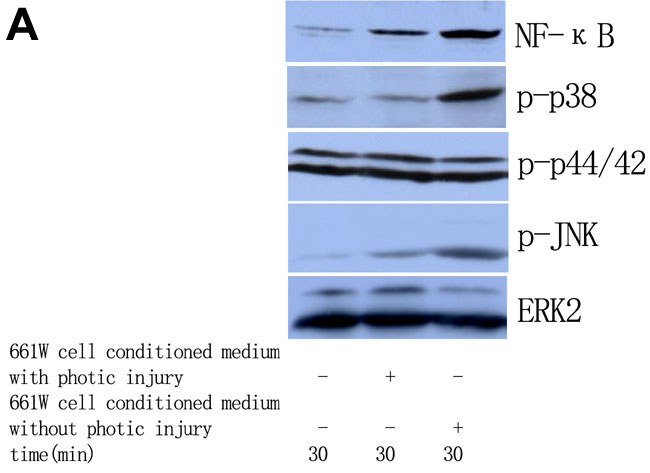
Figure 7. The effect of photoreceptor cell conditioned medium on NF-κB p65 levels and MAPK phosphorylation in retinal microglia
A: Representative western blots images showed the time course of changes in levels of NF-κB p65, phosphorylated p38 (p-p38), phosphorylated p44/42 (p-p44/42), and phosphorylated JNK (p-JNK) in response to photoreceptor cell conditioned medium with or without photic injury. B: Quantified levels of the indicated protein. The relative levels of each protein were normalized to ERK2. *p<0.01 versus cells grown in basal medium (NC). Hash mark p<0.01 versus cells grown in 661W cell conditioned medium without photic injury.