![]() Figure 3 of
Yang, Mol Vis 2007;
13:2048-2057.
Figure 3 of
Yang, Mol Vis 2007;
13:2048-2057.
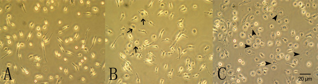
Figure 3. The effect of photoreceptor cell conditioned medium on retinal microglial morphology
A: Phase contrast images of primary retinal microglial cells cultured in basal media for 48 h. B: Following exposure to 661W cell conditioned medium without photic injury, some cells became larger and rounder (arrow), some cells remain unchanged. C: Following exposure to 661W cell conditioned medium with photic injury (PRCM), the cells grew notably the characteristic ameboid shape of activated microglia (arrowhead).