![]() Figure 1 of
Scholl, Mol Vis 2007;
13:196-205.
Figure 1 of
Scholl, Mol Vis 2007;
13:196-205.
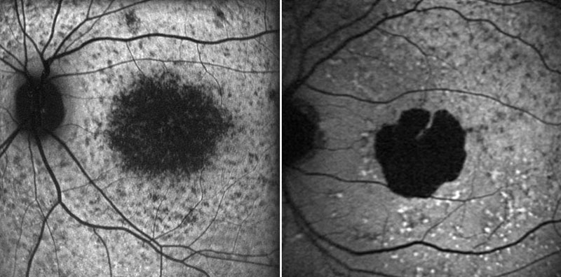
Figure 1. Phenotyping by means of fundus autofluorescence imaging
Fundus autofluorescence images obtained with a cSLO (Heidelberg retina angiograph, HRA 2, Heidelberg Engineering, Dossenheim, Germany) according to a standard operating procedure. Left: Patient diagnosed with Stargardt's macular dystrophy (age, 17 years); right: patient diagnosed with atrophic AMD (GA) and a fundus autofluorescence pattern "diffuse-fine granular with peripheral punctate spots" according to Bindewald et al. [34] (age: 71 years).