![]() Figure 2 of
Hansen, Mol Vis 2007;
13:2019-2022.
Figure 2 of
Hansen, Mol Vis 2007;
13:2019-2022.
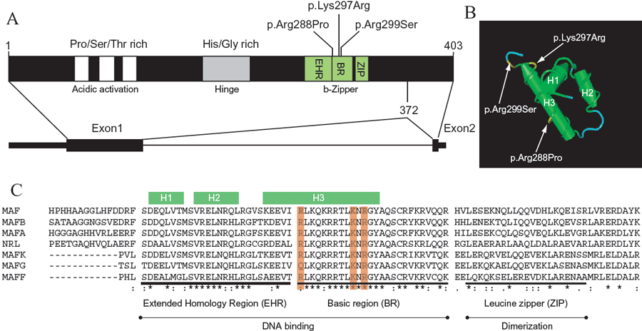
Figure 2. Structure of the human MAF protein and gene
A illustrates the schematic representation of the MAF protein and gene structure. The three known mutations in the bZip region are indicated. B shows a structural analysis of MafG showing three α-helixes in the bZIP region. The 3D-structure is based on PDB data: 1K1V [17]. All three CCMC mutations involved in CCMC are interfering with helix 3. C shows that the protein sequence alignment of the seven human MAF proteins reveals extreme high conservation in the EHR and BR element. (The protein accession numbers are: Maf, NP_005351; MafA NP_963883; MafB, NP_005452; MafF, NP_690617; MafK, NP_002351; MafG, NP_002350; Nrl, NP_006168).