![]() Figure 9 of
McGowan, Mol Vis 2007;
13:1984-2000.
Figure 9 of
McGowan, Mol Vis 2007;
13:1984-2000.
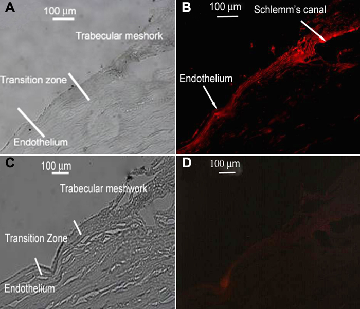
Figure 9. Wnt-1 in the wounded cornea
Cornea is from a 50-year-old male. Brightfield image (A) and corresponding fluorescent image (B) showing Wnt-1 presence in the trabecular meshwork, particularly the endothelial border of Schlemm's canal of a wounded cornea. Wnt-1 is not present in the trabecular meshwork of a 38-year-old whose cornea was unwounded. (C, D). All images are cross sections. Control slides showed no reaction (not shown).