![]() Figure 6 of
McGowan, Mol Vis 2007;
13:1984-2000.
Figure 6 of
McGowan, Mol Vis 2007;
13:1984-2000.
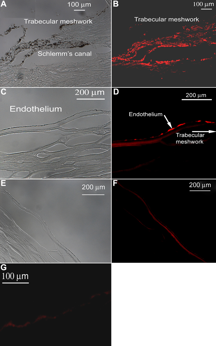
Figure 6. Telomerase in the trabecular meshwork and peripheral corneal endothelium
The cornea is from a 50-year-old male. Brightfield images (A, C) and corresponding fluorescent images show the presence of telomerase in the trabecular meshwork (B) and the peripheral corneal endothelium (D) of an unwounded cornea. Telomerase was not present in the central endothelium (E) although there is some autofluorescence indicated in the stroma at the border of Descemet's membrane(F). The staining pattern was identical in the wounded sections (not shown). Control slides showed no reaction (G).