![]() Figure 4 of
McGowan, Mol Vis 2007;
13:1984-2000.
Figure 4 of
McGowan, Mol Vis 2007;
13:1984-2000.
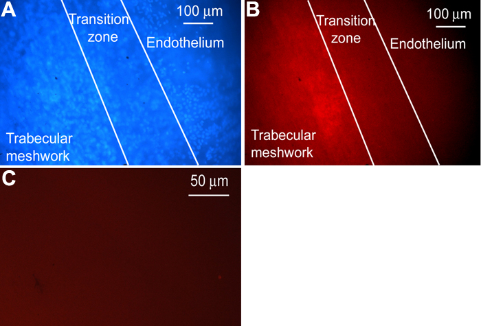
Figure 4. Nestin in the wounded cornea
The cornea is from a 44-year-old. The DAPI image (A) and fluorescent image (B) in the wounded cornea show nestin in the trabecular meshwork and the adjacent transition zone. The DAPI image shows cell nuclei. Control slides showed no reaction (not shown).