![]() Figure 2 of
McGowan, Mol Vis 2007;
13:1984-2000.
Figure 2 of
McGowan, Mol Vis 2007;
13:1984-2000.
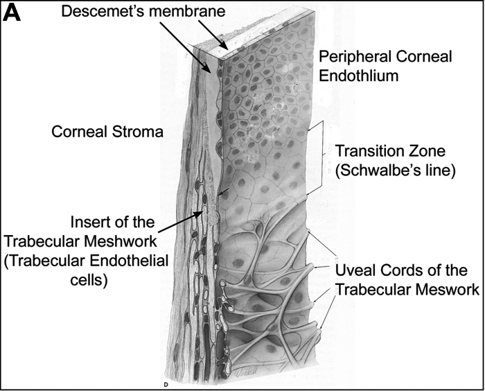
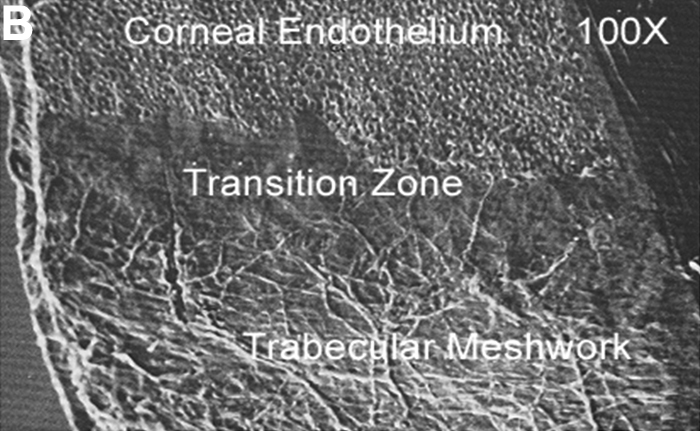
Figure 2. Three-quarter and flat mount views of the human cornea and limbus
The diagram at the top (A) illustrates the anatomic relationships of the tissues and regions discussed in this study. The area indicating the insert of the trabecular meshwork includes cells at the border of Schlemm's canal. A scanning electron micrograph of a human corneal flat mount is shown in B. Adapted from Histology of the Human Eye, Hogan, Alvarado, Weddell, The Limbus, p. 176-177, 1971, with permission from Elsevier.