![]() Figure 10 of
McGowan, Mol Vis 2007;
13:1984-2000.
Figure 10 of
McGowan, Mol Vis 2007;
13:1984-2000.
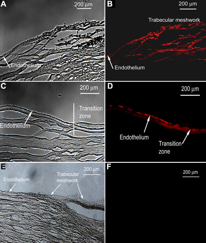
Figure 10. Pax-6 in the wounded cornea
The cornea is from a 38-year-old male. Brightfield images (A, C) and corresponding fluorescent images (B, D) show the presence of Pax-6 in the trabecular meshwork and the corneal endothelium of a wounded cornea. Pax-6 is not present in unwounded tissue from a 29-year-old male (E, F). All images are cross sections. Control slide showed no reaction (not shown).