![]() Figure 1 of
McGowan, Mol Vis 2007;
13:1984-2000.
Figure 1 of
McGowan, Mol Vis 2007;
13:1984-2000.
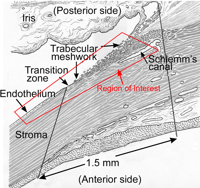
Figure 1. Transverse diagram of the human cornea and limbus
The diagram illustrates the anatomic relationships of the tissues and regions discussed in this study (outlined in red). The posterior limbus is the region between the solid vertical lines as shown, consisting of the transition zone, the trabecular meshwork, and Schlemm's canal. The corneal endothelium is not part of the limbus. Adapted from Histology of the Human Eye, Hogan, Alvarado, Weddell, The Limbus, p.113, 1971, with permission from Elsevier.