![]() Figure 3 of
Takacs, Mol Vis 2007;
13:1976-1983.
Figure 3 of
Takacs, Mol Vis 2007;
13:1976-1983.
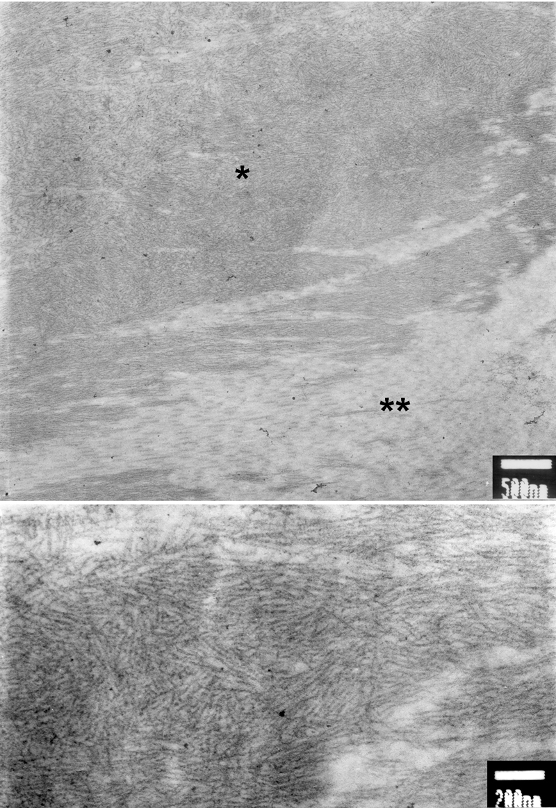
Figure 3. Electron micrograph of the cornea with the F547S mutation
Top: Electron micrographs of the cornea of the patient with the F547S mutation shows a large, electron-dense deposit (asterisk) among the collagen lamellae (double asterisk). Bottom: At higher magnification, nonbranching 8-10 nm fibrils are revealed, indicating the amyloid nature of the deposit.