![]() Figure 2 of
Takacs, Mol Vis 2007;
13:1976-1983.
Figure 2 of
Takacs, Mol Vis 2007;
13:1976-1983.
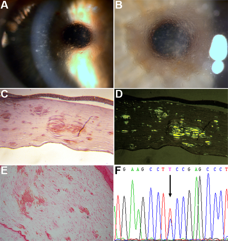
Figure 2. Slit lamp and histological images of the polymorph corneal amyloidosis caused by the F547S mutation
A and B: Clinical pictures of the polymorph corneal amyloidosis caused by the F547S mutation shows a snowflake-like central and fine linear corneal deposits. Deposits extend to the pre-Descemet level as shown in the photography with narrow slit (A). C: Congo red-stained cornea of the same patient shows large congophilic deposits in the corneal stroma, which exhibit green birefringence when viewed under polarized light (D). E: Cornea of the same patient, stained with anti-BIGH3 antibody, is shown in this section. All deposits are stained in red. F: The electropherogram of exon 12 of TGFBI of the same patient is shown. The arrow indicates the T>C heterozygous conversion at position 1640, causing the F547S amino acid exchange.