![]() Figure 1 of
Takacs, Mol Vis 2007;
13:1976-1983.
Figure 1 of
Takacs, Mol Vis 2007;
13:1976-1983.
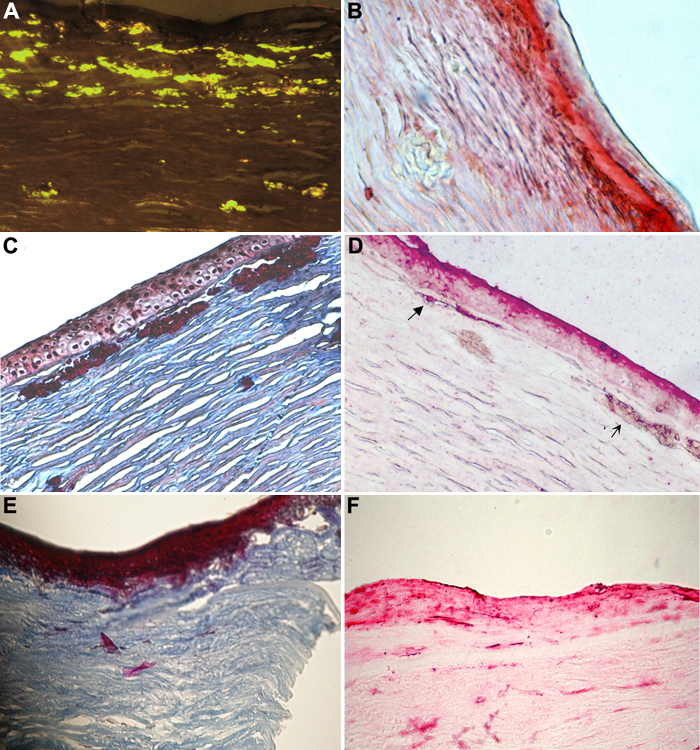
Figure 1. Histological examination of BIGH3-related corneal dystrophies
A: A congo red-stained LCDI cornea is shown under polarized light. Amyloid deposits are light green. B: Immuno-histological staining of an LCDI cornea with anti-BIGH3 antiserum is shown. Intra- and subepithelial deposits show red staining. C: In a GCDI cornea, deposits appear bright red with Masson's trichrome stain. D: Immuno-histological labeling of a cornea with recurrent GCDI shows the presence of BIGH3 protein in the deposits. E and F: Masson's trichrome and immuno-histological staining of a recurrent Thiel-Behnke dystrophy is illustrated. Subepithelially deposited fibrous material stains blue with Masson's trichrome and shows intensive immuno-labeling with the anti-BIGH3 antibody. Note that in E the epithelium is missing.