![]() Figure 1 of
Rosenberg, Mol Vis 2007;
13:1962-1969.
Figure 1 of
Rosenberg, Mol Vis 2007;
13:1962-1969.
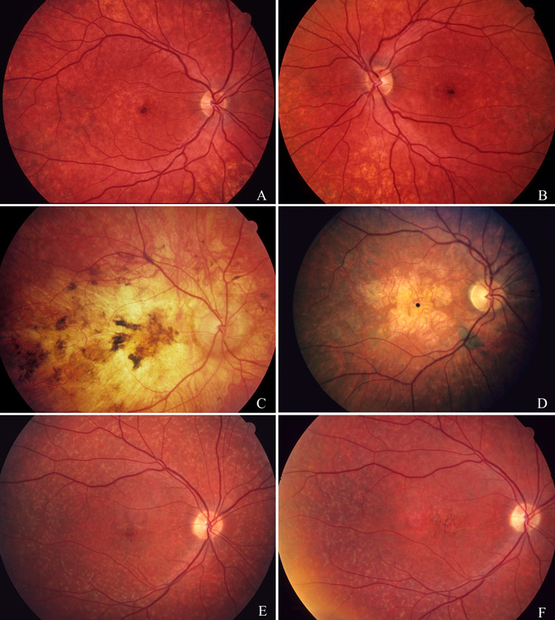
Figure 1. Fundus pictures of four patients homozygous for N965S showing stages of different ABCA4 phenotypes
A and B: Bull's eye phenotype is evident in D065, age 17 years old. In the photograph, taken four years after onset of the phenotype, the posterior poles in right and left eyes, show inconspicuous, ill-defined, spotty atrophies in the macular region and peripherally. No yellow spots were present. C: Diffuse choroidoretinal dystrophy phenotype in D137, age 48 years old. In the photograph taken 30 years after onset, a total atrophy of the choriocapillaris-retinal pigment epithelial layers occupies the macular area with peripapillary extensions. Coarse central pigment aggregates and ill-defined margins further characterize the picture. D: Cone-rod phenotype in D174, 32 years old. In the photograph taken 19 years after onset, there is an irregular geographic atrophy surrounded by a zone of atrophy of the choriocapillary layer. No white spots are seen at this late stage. E: D199, age 19 years, showing Stargardt-flavimaculatus phenotype at four years after onset. E and F: Fundus photograph of D199, age 19 and 22 years, taken at four and seven years after onset. In both photographs, there are midperipheral, small, yellow-gray spots that surround an indistinct atrophy of the macular pigment epithelium. A progression of the central RPE atrophy occurred during the three year intervals.