![]() Figure 3 of
Dentchev, Mol Vis 2007;
13:190-195.
Figure 3 of
Dentchev, Mol Vis 2007;
13:190-195.
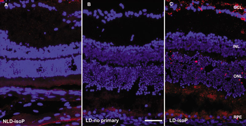
Figure 3. Fluorescence photomicrographs of Balb/c mouse retinas
A: Normal retina, no bright light exposure, labeled with anti-isoprostane antibody (red fluorescence). B: Control retina without primary antibody after 28 h bright light exposure. C: Retina after 28 h bright light labeled with anti-isoprostane antibody exposure. Nuclei are labeled with DAPI (blue). Scale bar indicates 50 μm. Abbreviations are defined as follows: no light damage (NLD), light damage (LD), exposure to anti-isoprostane antibody (isoP).