![]() Figure 7 of
Kim, Mol Vis 2007;
13:1942-1952.
Figure 7 of
Kim, Mol Vis 2007;
13:1942-1952.
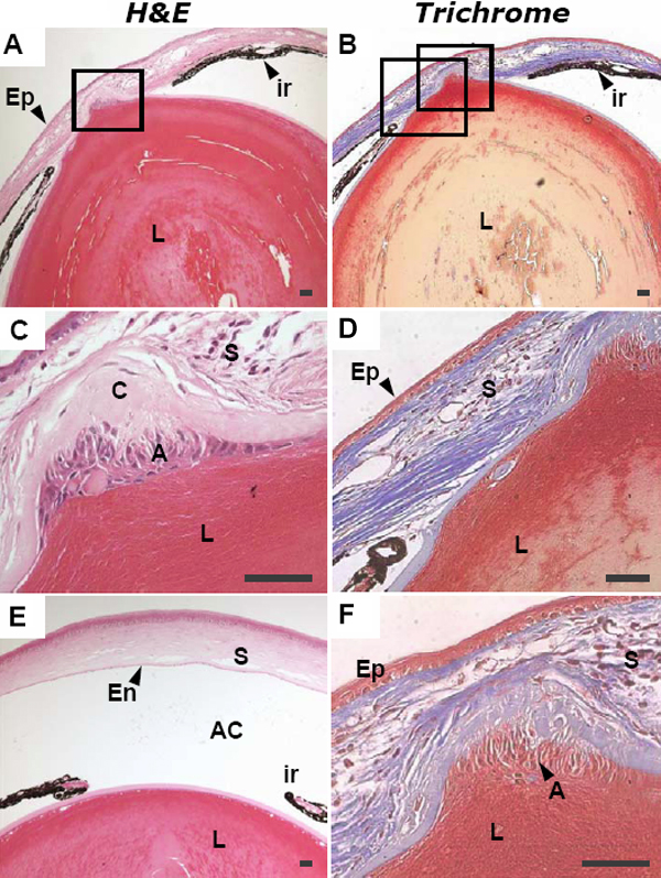
Figure 7. Histologic analysis of the anterior segment in a seven-month-old Alb-hβigh3 transgenic mouse
Staining was performed with H&E (A, C, and E) and Trichrome (B, D, and F) in each 4 μm thick paraffin section. Images in rows 2 and 3 are higher-magnification images corresponding to the small rectangles in column 1. Separation between lens and cornea was not complete in the defective eye of the Alb-hβigh3 transgenic mouse. The attached portion between the protruded lens and cornea showed proliferated lens epithelial cell under the lens capsule. The terminal part of the iris was attached to the posterior surface of the cornea and there was no space for the anterior chamber. E shows the normal eye of a seven-month-old wild-type mouse. The scale bars are equal to 50 μm. Abbreviations: L, Lens; C, lens capsule; A, anterior epithelium of lens; ir, iris; Ep, corneal epithelium; S, corneal stroma; En, corneal endothelium.