![]() Figure 6 of
Kim, Mol Vis 2007;
13:1942-1952.
Figure 6 of
Kim, Mol Vis 2007;
13:1942-1952.
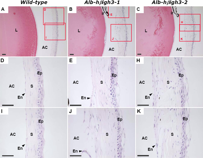
Figure 6. Histological examination of the defective eye in transgenic mouse compared to normal eye in wild-type mouse at two months of age
Staining was performed with H&E in each 4 μm thick paraffin sections. C is the serial section of B. Images in rows 2 and 3 are higher-magnification images of the cornea corresponding to the two red rectangles in column 1. The abnormal cornea of transgenic mice (B, C) had disorganized corneal stroma and disconnected Descemet's membrane and endothelial cell layer compared to wild-type (A). The scale bars are equal to 50 μm. Abbreviations: L, Lens; ir, iris; AC, anterior chamber; Ep, corneal epithelium; S, corneal stroma; En, corneal endothelium.
