![]() Figure 5 of
Kim, Mol Vis 2007;
13:1942-1952.
Figure 5 of
Kim, Mol Vis 2007;
13:1942-1952.
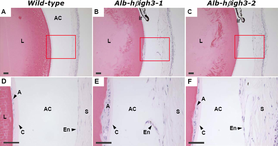
Figure 5. Histopathologic findings of normal eye in wild-type mouse and the defective eye in transgenic mouse at two months of age
Staining was performed with H&E in each 4 μm thick paraffin section. C is the serial section of B. Images in row 2 are higher-magnification images corresponding to small red rectangles in row 1. The abnormal cornea of transgenic mice (B, C) had disorganized corneal stroma, disconnected corneal endothelium, and a narrow anterior chamber compared to wild-type (A). The anterior surface of the lens had multilayer epithelial cells in transgenic mice. The scale bars are equal to 50 μm. Abbreviations: L, lens; C, lens capsule; A, anterior epithelium of lens; AC, anterior chamber; ir, iris; S, corneal stroma; En, corneal endothelium.