![]() Figure 4 of
Kim, Mol Vis 2007;
13:1942-1952.
Figure 4 of
Kim, Mol Vis 2007;
13:1942-1952.
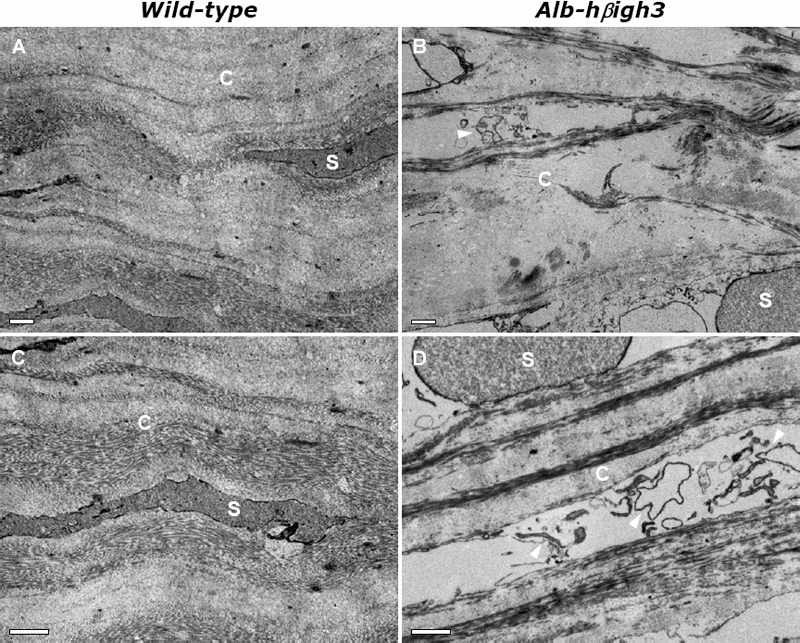
Figure 4. Electron micrographs showing stroma of central portion of cornea in wild-type and Alb-hβigh3 transgenic mice
Corneas from mice were fixed in 2.5% glutaraldehyde and subjected to electron microscopy. A and C: In wild-type mice, collagen fibers and fibrils were regularly and compactly arranged parallel to the epithelial surface. However, in Alb-hβigh3 transgenic mice (B and D), the collagen fibers and fibrils were disorganized. Tissue debris was occasionally observed between the collagen fibers (arrowhead). A and B are images corresponding to 5K and C and D are images corresponding to 8K. The scale bars are equal to 2 μm. Abbreviations: C, collagen layers; S, stromal cells.