![]() Figure 2 of
Qi, Mol Vis 2007;
13:1934-1941.
Figure 2 of
Qi, Mol Vis 2007;
13:1934-1941.
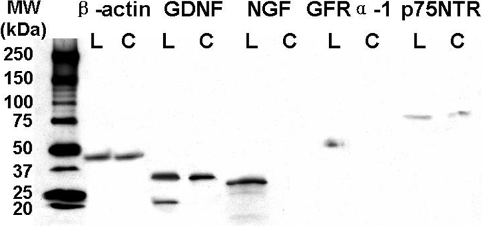
Figure 2. Western blot analysis
Western blot analysis of GDNF, NGF, GFRα-1, and p75NTR in human corneal (C) and limbal (L) epithelial tissues with β-actin (45 kDa) as a control. NGF (a major band at approximately 30 kDa, and two weaker bands at 25 and 14 kDa) and GFRα-1 (53 kDa) were detected only in the limbal epithelium and not in the cornea. A 35 kDa band of glycosylated GDNF was detected in both limbal and corneal epithelia while an additional 21 kDa band was only detected in limbal epithelium. The p75NTR protein (75 kDa) was detected in both limbal and corneal epithelia.