![]() Figure 1 of
Qi, Mol Vis 2007;
13:1934-1941.
Figure 1 of
Qi, Mol Vis 2007;
13:1934-1941.
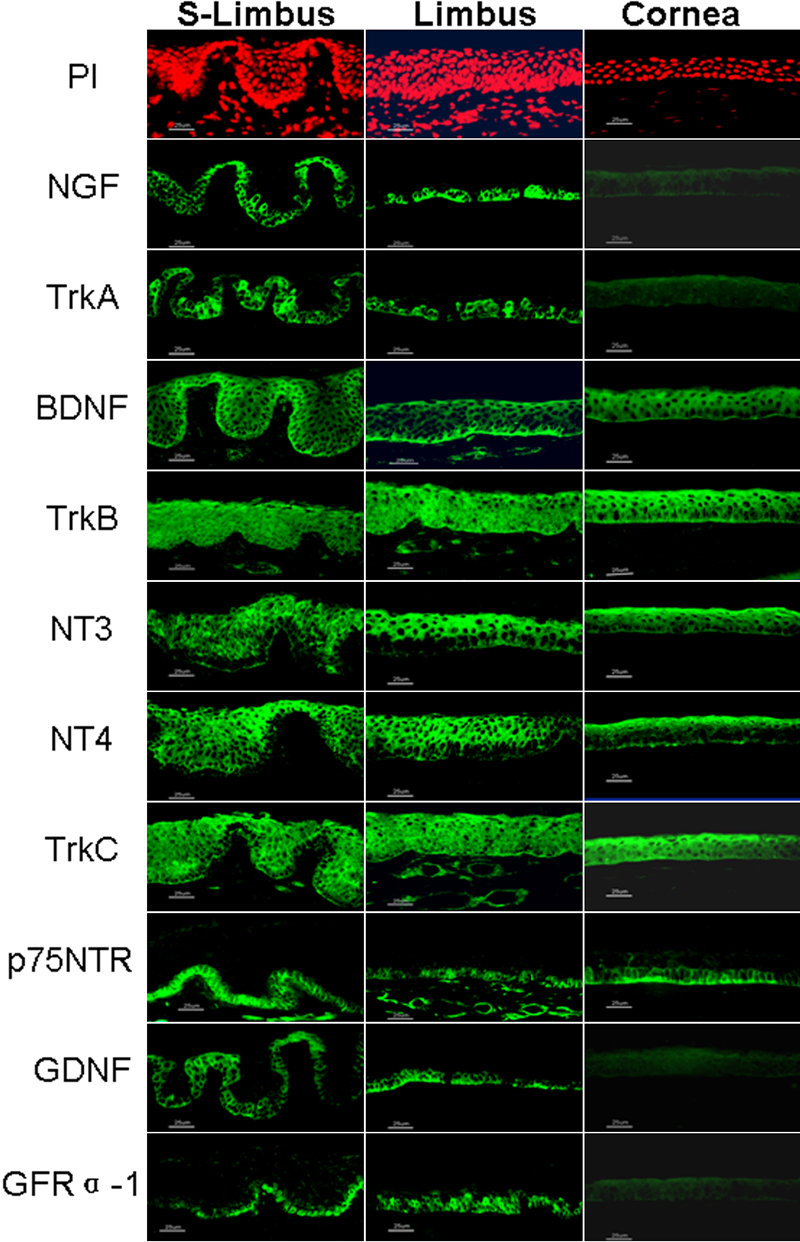
Figure 1. Expression of neurotrophic factors and corresponding receptors in human corneal tissue
Representative images showing the immunofluorescent staining of NGF, TrkA, BDNF, TrkB, NT-3, NT-4, TrkC, p75NTR, GDNF, and GFRα-1, (green color) in frozen sections of human corneoscleral tissues: tangential cross sections cut through the superior limbus (S-Limbus, left column), radial meridional sections cut through limbus (Limbus, central column), and central cornea (Cornea, right column). Propidium iodide (PI) was used as nuclear counterstaining (red). The images in the top panel are matched to the images in the NGF panel as examples; other PI images matched to each NTF staining are not shown. Magnification: x400 (bar = 25 μm).