![]() Figure 3 of
Chow, Mol Vis 2007;
13:1926-1933.
Figure 3 of
Chow, Mol Vis 2007;
13:1926-1933.
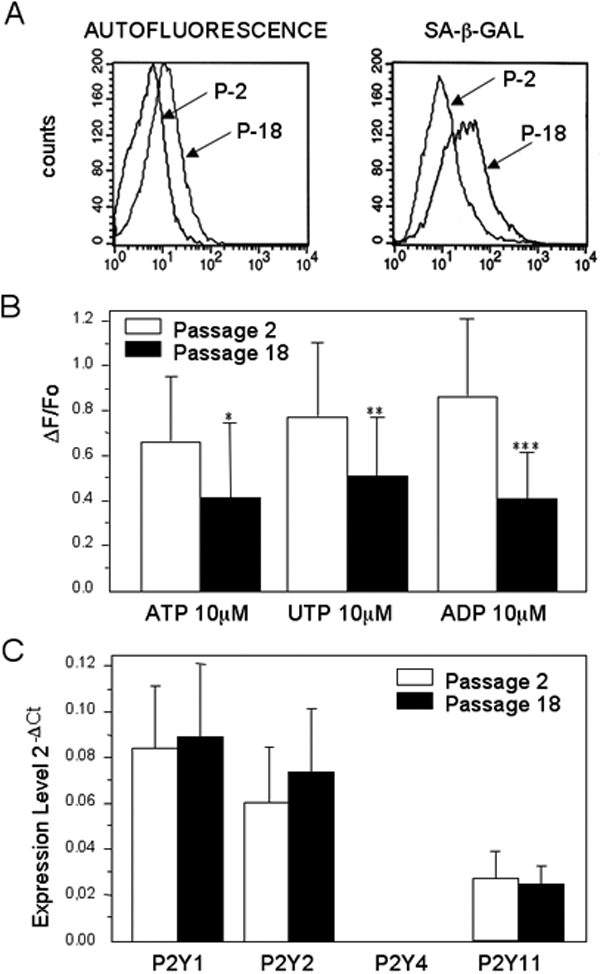
Figure 3. Effect of replicative senescence on P2Y receptor expression and function
The increase of the cellular senescence markers, autofluorescence, and sa-β-gal activity in cells at passage 18 (P-18) compared to cells at passage 2 (P-2) was evaluated by FACS analysis (A). The data are representative of three independent experiments. To evaluate whether cellular senescence leads to a decrease in the ability of the TM cells to respond to P2Y agonists, the calcium responses (ΔF/F0) to all three agonists were measured in TM cells at passage 18 and compared with cells at passage 2. The decline in ΔF/F0 in cells at passage 18 was statistically significant in all cases (The asterisk indicates p=0.03, n=15; the double asterisk means p<0.001, n=30; three asterisks indicate that p=0.001, n=12; B). The decrease in ΔF/F0 was not associated with any significant decrease in expression of the mRNAs for P2Y1, P2Y2, and P2Y11. The mRNA for P2Y4 receptor subtype was not detected in TM cells. The functionality of our designed primer was verified with signal isolation from a genomic DNA sample. The P2Y11 subtype appeared at a much later PCR cycle, suggesting that it is present in smaller quantities than P2Y1 and P2Y2 receptor subtypes (C).