![]() Figure 3 of
Bernstein, Mol Vis 2007;
13:1920-1925.
Figure 3 of
Bernstein, Mol Vis 2007;
13:1920-1925.
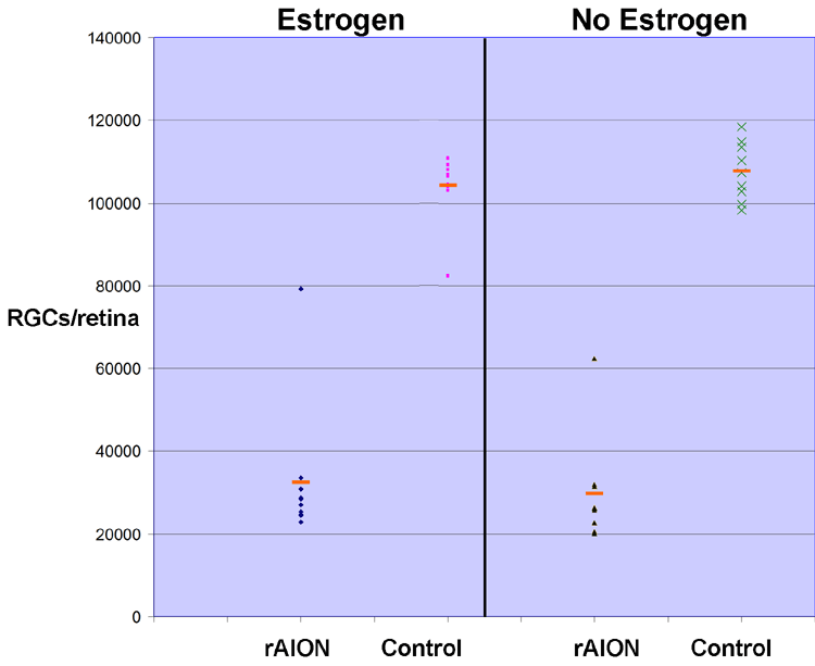
Figure 3. Stereological analysis after rodent anterior ischemic optic neuropathy induction retinal ganglion cell loss in estrogen- and vehicle-treated animals
Immediately after rodent anterior ischemic optic neuropathy (rAION) induction, animals received an intramuscular injection of either 50 μg/kg 17-estradiol, or vehicle (sesame oil) as described in Methods. Retinal ganglion cells (RGCs) were retrograde-labeled with fluorogold, and animals euthanized 28 days post-induction. Retinas were post-fixed, flat mounted and analyzed as described in Methods. A 65% average RGC loss is seen in estrogen treated animals, compared with a 70% RGC loss in vehicle treated animals. Control eyes: contralateral (naíve) eyes from each treatment group. Red bars indicate the mean RGC count for each group.
