![]() Figure 1 of
Bernstein, Mol Vis 2007;
13:1920-1925.
Figure 1 of
Bernstein, Mol Vis 2007;
13:1920-1925.
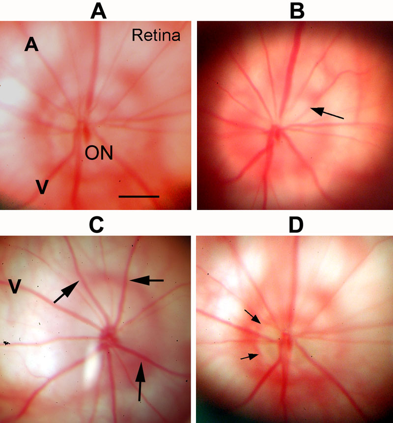
Figure 1. Appearance of estrogen- and vehicle treated retina and optic nerve before and after induction of rodent anterior ischemic optic neuropathy
A: Pre-induction. The retina (Retina), optic nerve (ON) and retinal vessels (A and V) are normal in appearance. B: Same eye as A two days after-rodent anterior ischemic optic neuropathy (rAION) and estrogen treatment. The ON disk margin is blurred and edematous (arrow). C: rAION-induced+, vehicle-treated eye, two days post-induction. The ON is edematous, demonstrable as an alteration in direction of the retinal vessels emerging from the ON (arrows). D: ON appearance in estrogen treated animal 21 days post-rAION induction. The ON disk margin is apparently shrunken, with marked pallor (arrows). The scale bar represents 250 μm.