![]() Figure 1 of
Miyazawa, Mol Vis 2007;
13:1912-1919.
Figure 1 of
Miyazawa, Mol Vis 2007;
13:1912-1919.
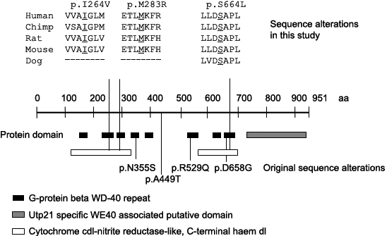
Figure 1. Multiple amino acid alignment and evolutionary conservation of p.I264, p.M283, and p.S664.
Protein domains of WDR36 are shown, and reported missense mutations are described below the schematic structure. p.I264, p.M283, and p.S664 are conserved among four (five) species and are located in the second, third, and eighth G-beta WD40 repeat as well as the Cytochrome cd1-nitrite reductase-like, COOH-terminal haem d1 (cyt cd1) domain, respectively.