![]() Figure 5 of
Wajchman, Mol Vis 2007;
13:1902-1911.
Figure 5 of
Wajchman, Mol Vis 2007;
13:1902-1911.
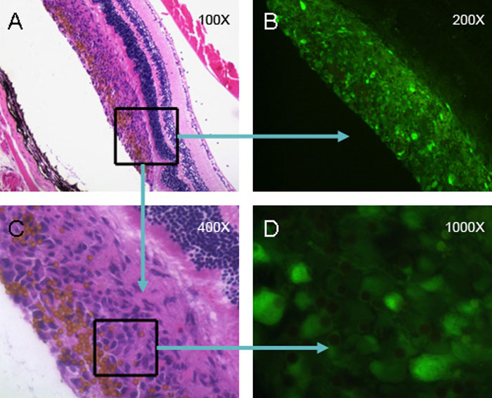
Figure 5. Delivery of retinal pigment epithelium and microbeads to the subretinal space
Retinal pigment epithelium from B6.GFP mice were mixed with an equal number of microbeads (shown as small, orange spheres) and injected into the subretinal space of B6 mice. The mice were euthanized the next day and their eyes sectioned and stained with H&E and examined by light microscopy (A and C) or fluorescent microscopy (B and D).