![]() Figure 4 of
Wajchman, Mol Vis 2007;
13:1902-1911.
Figure 4 of
Wajchman, Mol Vis 2007;
13:1902-1911.
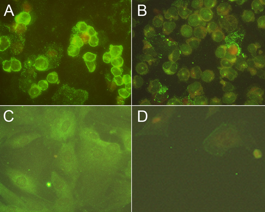
Figure 4. Expression of the ovalbumin protein in transduced cells
Expression of the OVA protein was detected by staining slides with a rabbit anti-OVA antibody followed by FITC-labeled goat anti-rabbit antibody and counter-staining with Evan's blue. Oovalbumin expressing E.G7-OVA cells (A) are compared to EL4 (B) as positive and negative controls, respectively. Retinal pigment epithelium (RPE) from B6 TRP-1-OVA mice (C) are compared to RPE from B6 mice (D).