![]() Figure 2 of
Wajchman, Mol Vis 2007;
13:1902-1911.
Figure 2 of
Wajchman, Mol Vis 2007;
13:1902-1911.
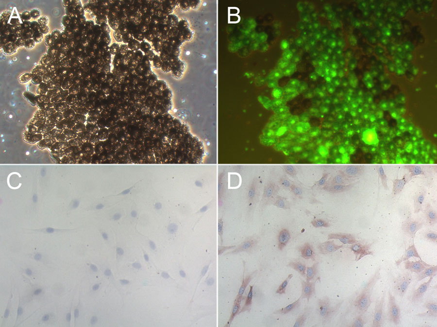
Figure 2. Isolation and identification of retinal pigment epithelium
Freshly isolated retinal pigment epithelium from B6.GFP mice demonstrate a typical hexagonal pattern of pigmented cells shown by dark field microscopy (A), virtually all of which were fluorescent (B). These RPE were cultured for 2 weeks and stained with an isotype control antibody (C) or anti-cytokeratin 8 (D) which showed cytoplasmic staining verifying their epithelial origin.