![]() Figure 3 of
Siegwart, Mol Vis 2007;
13:1878-1886.
Figure 3 of
Siegwart, Mol Vis 2007;
13:1878-1886.
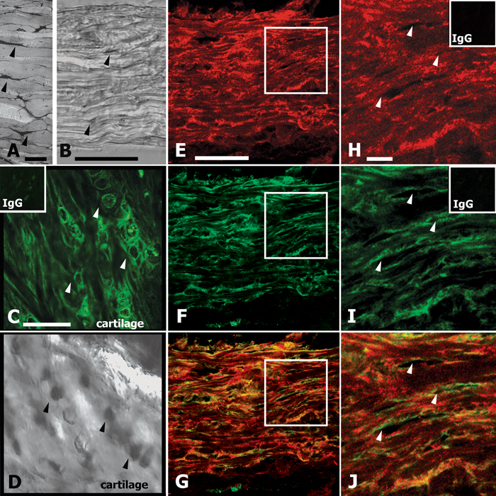
Figure 3. Immunohistochemical localization of type I collagen and aggrecan in tree shrew sclera
A and B: Cross sectional views of tree shrew sclera are shown. A: Electron micrograph is stained with cuprolinic blue (scale=10 μm, photograph by Edward Clarke). The individual lamellae can be seen with fibroblast cell bodies and processes (black structures, examples marked with arrowheads) between the lamellae. B: Differential interference contrast (DIC) image is counterstained with Quickstain (scale=50 μm). E-J: Single optical sections (400 nm thickness) show type I collagen immunoreactivity (E, scale=50 μm and H, enlargement of the area indicated by the box in E, scale=10 μm), aggrecan immunoreactivity (F and I), and overlay images (G and J). Although the intensity varies, the type I collagen immunoreactivity is essentially continuous throughout the sclera while aggrecan immunoreactivity is less continuous and appears restricted to thin bands. Overlay images (G and J) show that there are areas of discrete collagen labeling (red) and discrete aggrecan labeling (green) as well as areas where collagen and aggrecan appear to colocalize in thin bands (yellow). Voids with no collagen or aggrecan labeling (arrows in H, I, and J) are likely where the thin optical section passes through the interior of fibroblast bodies and processes. This can be seen most clearly in J where rings of discrete aggrecan immunoreactivity appear to surround the regions with no immunoreactivity (arrowheads). C and D: Aggrecan immunoreactivity in cartilage served as a positive control. There is diffuse immunoreactivity throughout the cartilage as well as more intense immunoreactivity surrounding the presumptive chondrocytes (C; examples marked with arrowheads; scale = 50 μm). DIC images of a comparable counterstained section (D) supports that the intense circular aggrecan immunoreactivity is likely at the surface of the round chondrocytes. No immunoreactivity was seen in negative controls substituting matched concentrations of rabbit or goat IgG for the respective primary antibody (insets in C, H, and I).