![]() Figure 1 of
Zhang, Mol Vis 2007;
13:1873-1877.
Figure 1 of
Zhang, Mol Vis 2007;
13:1873-1877.
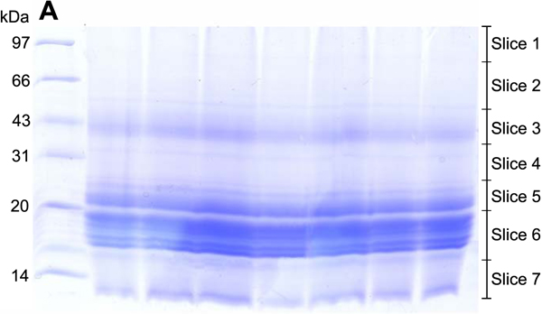
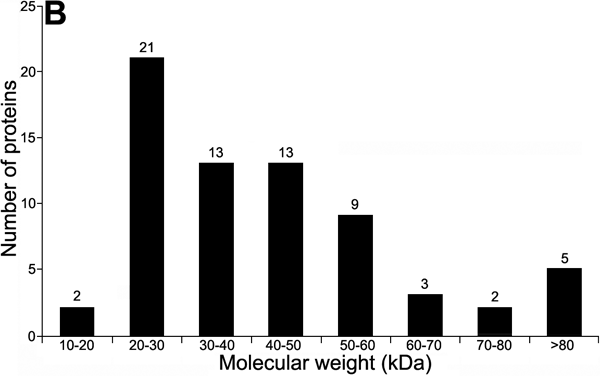
Figure 1. SDS-PAGE separation of lens proteins and the distribution of theoretical molecular weights of the identified proteins
A: The proteins isolated from epithelium-free human lens fibers were resolved by non-continuous SDS-PAGE. Thirty-two micrograms of protein mixture was loaded onto each lane. The first lane on the right shows the markers of molecular weights. The gels were stained with Coomassie brilliant blue cut into seven slices as indicated. Proteins in each slice of the gels were in-gel digested with trypsin, and the peptides were extracted and analyzed by RPLC-/MS/MS. B: The distribution of theoretical molecular weights of the identified proteins is shown.