![]() Figure 6 of
Pan, Mol Vis 2007;
13:181-189.
Figure 6 of
Pan, Mol Vis 2007;
13:181-189.
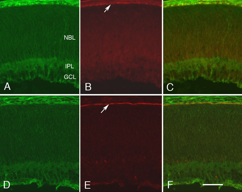
Figure 6. Colocalization of CD81 with Sap97 and EBP50 at P2
The staining pattern of CD81, Sap97, and EBP50 in the developing rat retina is shown at P2. A and D are stained for CD81, B is stained for Sap97, and C is the merged image. E is stained for EBP50; F is the merged image. At this age, the ganglion cell layer (GCL) and inner plexiform layer (IPL) are present. The remainder of the retina consists of a neuroblastic layer (NBL). The arrows in B and E indicate the location of the developing retinal pigment epithelium (RPE) layer. All photomicrographs were taken at the same magnification. The scale bar in represents 25 μm.