![]() Figure 5 of
Pan, Mol Vis 2007;
13:181-189.
Figure 5 of
Pan, Mol Vis 2007;
13:181-189.
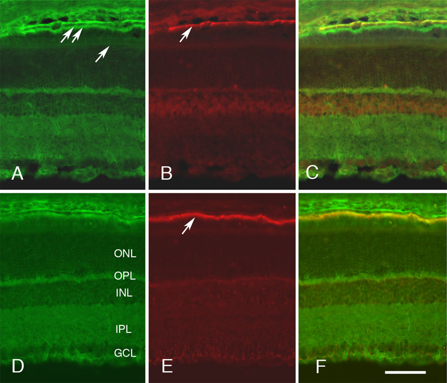
Figure 5. Colocalization of CD81 with Sap97 and EBP50 at P20
The staining of the P20 rat retina for CD81 (A and D), Sap97 (B), and EBP50 (E) is shown in two double-stained sections. The merged images of these two sections are shown in C and F. The pattern of CD81 labeling is consistent with the labeling of Müller glial cells, as shown by the prominent labeling of the external limiting membrane (arrow, A) and retinal pigment epithelium (RPE) cells (double arrow, A). Sap97 shows a general stain in the retinal neurons and a prominent band at the base of the RPE (arrow in B). In the merged image (C), Sap97 immunoreactivity is colocalized with CD81 on the basolateral surface of RPE cells. EBP50 labels a prominent band at the junction between the retina and the PRE cells (arrow, E). The EBP50 immunoreactivity colocalizes with CD81 immunoreactivity at the apical surface of the RPE cells (F). In A and F, the layers of the P20 retina are shown: the ganglion cell layer (GCL), inner plexiform layer (IPL), inner nuclear layer (INL), outer plexiform layer (OPL), and outer nuclear layer (ONL). All photomicrographs are taken at the same magnification. The scale bar in F represents 25 μm.