![]() Figure 1 of
Spellicy, Mol Vis 2007;
13:1866-1872.
Figure 1 of
Spellicy, Mol Vis 2007;
13:1866-1872.
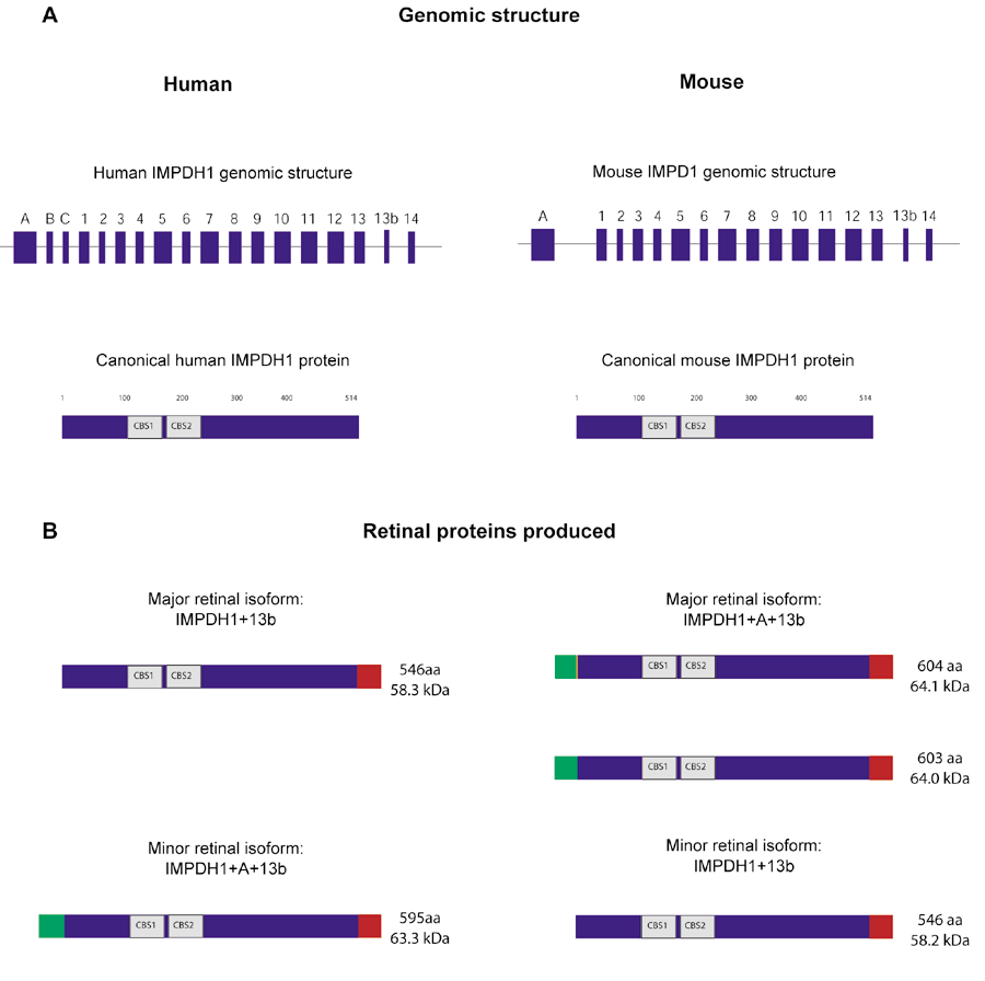
Figure 1. Human and mouse IMPDH1
A: The genomic structure of IMPDH1 in mouse and human, and the canonical IMPDH1 protein previously identified in human and mouse tissues [21]. B: The novel IMPDH1 protein isoforms recently identified in both human and mouse retinas. These retinal isoforms are produced through alternate starts of translation and alternate splicing leading to inclusion and/or exclusion of amino acid residues encoded by exons A and 13b. Green indicates exon A encoded residues and red indicates exon 13b encoded residues (adapted from Bowne et al, 2006) [18]. The two major retinal isoforms in mouse differ by only one amino acid (orange in figure) which results from alternate splicing of exon 1 adding 3 additional bases.
