![]() Figure 1 of
Boulouiz, Mol Vis 2007;
13:1862-1865.
Figure 1 of
Boulouiz, Mol Vis 2007;
13:1862-1865.
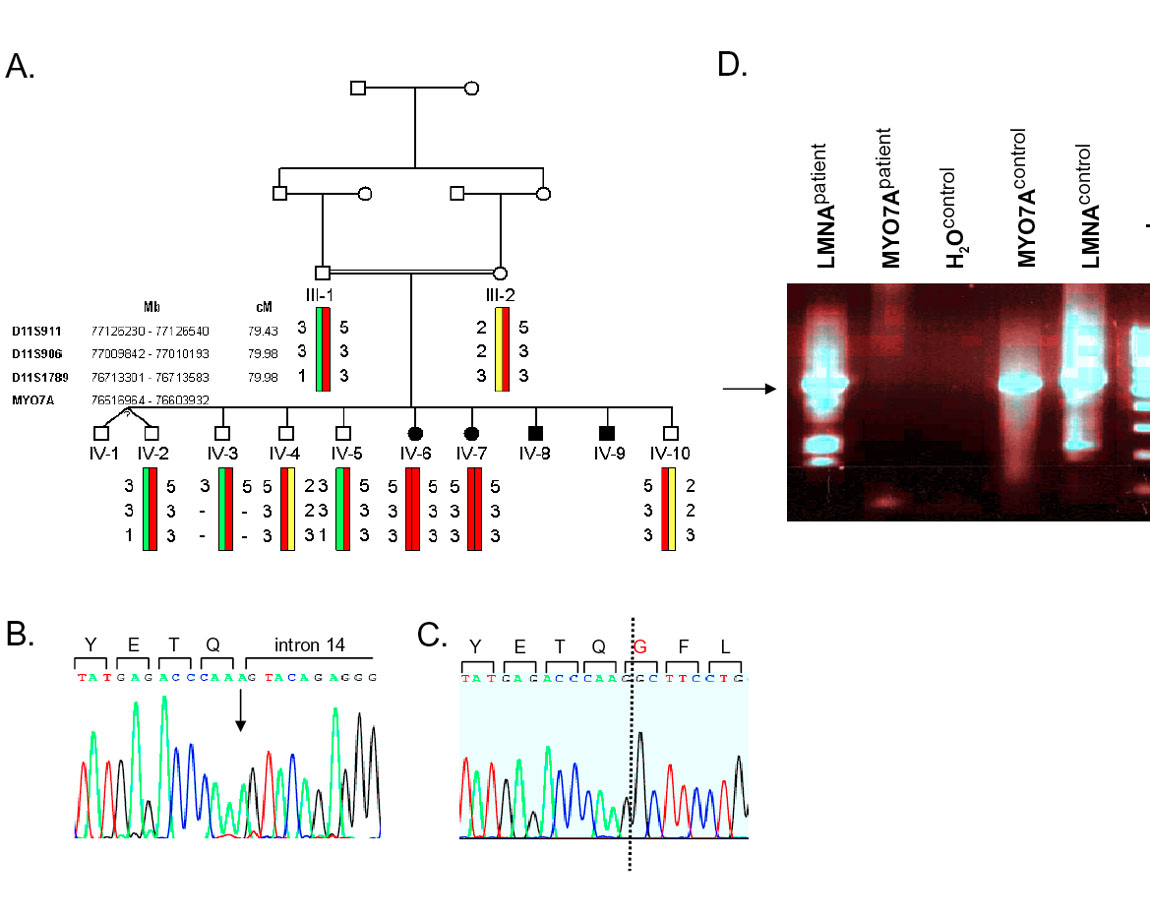
Figure 1. Pedigree and molecular studies of the Moroccan Usher syndrome type 1B family
A: Pedigree of the USH1B family and haplotypes of polymorphic markers in the MYO7A region. Circles represent females, squares males in the pedigree. Affected individuals are shown by filled symbols. The exact physical (Mb) and genetic (cM) location of markers and MYO7A gene are given. B: Sequence chromatogram of individual IV-6 showing the 3' end of exon 14 and exon-intron boundary. The arrow indicates the position of the homozygous c.1687G>A mutations. Encoded amino acids and the 5' end of intron 14 are marked. C: Chromatogram of a partial MYO7A transcript around the site of mutation amplified from control cDNA as shown in D. Dotted line marks the exon14/exon15 boundary. D: Amplification of a 517 bp fragment from exon 13-16 of MYO7A (marked by arrow) and a 631 bp control fragment from exon 6-10 of the LMNA gene on cDNA from the index patient and a control.