![]() Figure 2 of
Iannaccone, Mol Vis 2007;
13:1856-1861.
Figure 2 of
Iannaccone, Mol Vis 2007;
13:1856-1861.
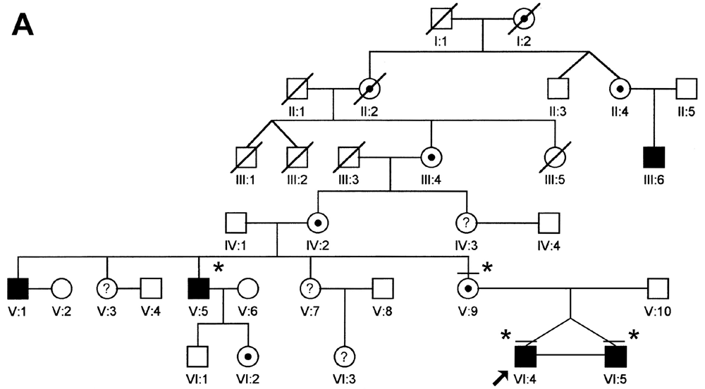
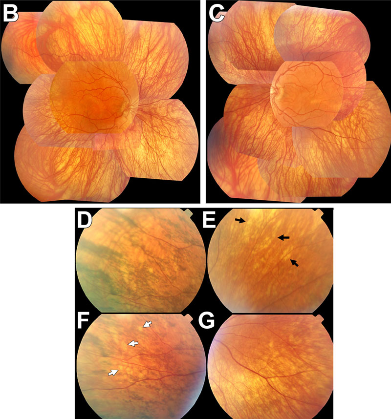
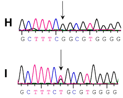
Figure 2. Midsouth family affected with X-linked ocular albinism
A: The pedigree of the midsouth family affected with X-linked ocular albinism. Asterisks mark represents subjects from whom samples were collected. The arrow points to the male proband analyzed. The bar above two of the symbols indicates the subjects were examined. Filled black symbols, denote affected males, and target round symbols indicate obligate or molecularly verified carriers. Question marks inside circles identify females whose status is unknown. Slashed symbols identify deceased subjects. B: A composite photograph of the fundus of the left eye of the proband (VI:4), a 15-year old boy with clinical signs of X-linked ocular albinism. C: A composite photograph of the fundus of the right eye of VI:5, the monozygotic twin of the proband, displaying findings similar to those of his brother (see text for details). Images D-G are representative fundus findings in V:9 (a 40 female carrier); D: A fundus image showing streaks of coarse, dark RPE mottling; E: A fundus image where black arrows mark a cluster of irregularly shaped streaks of depigmentation in an area of essentially normal fundus pigmentation; F: A fundus image where white arrows point to a cluster of nummular areas of depigmentation amid streaks of "mud-splattered" RPE mottling. G: An fundus image of an area of diffuse hypopigmentation in the infero-temporal retinal quadrant. H: OA1 sequence analysis from twin boys (VI:4 and VI:5) and of their maternal uncle (V:5). The arrow points to the 346T>G substitution, predicting a Cys116Arg amino acid change. I: Sequence analysis of V:9, showing T/G heterozygosity at position 346.