![]() Figure 4 of
Giordano, Mol Vis 2007;
13:1842-1850.
Figure 4 of
Giordano, Mol Vis 2007;
13:1842-1850.
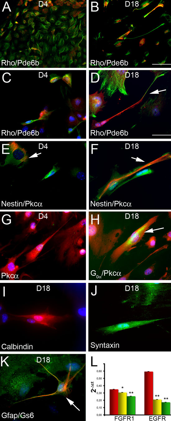
Figure 4. Differentiation into retinal-like neurons
Retinal stem cell were allowed to differentiate for four days (D4) or 18 days (D18) on an extracellular matrix (ECM) substrate. A-D: Rod-like cells were analyzed by co-localization of two different markers: Rhodopsin (Rho, green) and Pde6b (red). The arrow in D indicates a cell that co-expressed the two markers and in which Rho accumulated at the periphery of the cell. Scale bar in A and B represents 20 μm. Scale bar in D represents 10 μm as for all images from C-K. E-F: Progenitors and bipolar-like interneurons were distinguished by immunolabeling with antibodies anti-nestin (red) and anti-Pkcα (green). Progenitors co-expressed the markers nestin and Pkc-α (arrors). G-H: Bipolar-like cells were analyzed by co-localization of two different markers: Pkcα (red) and G0α (green). Arrow indicates a cell co-expressing the two markers after 18 days in differentiation. G0α was not expressed in cells after four days of differentiation. I: Immunolocalization of Calbindin, a marker for horizontal cells. J: Immunolocalization of Syntaxin, a marker for amacrine cells. K: Müller glia cells were analyzed by co-localization of two different markers: Gfap (red) and GS6 (green). Arrow indicates a cell that co-expressed the two markers. L: quantitative analysis of FGFR1 and EGFR mRNA expression in retinal progenitors cultured for 18 days in the presence of fibroblast growth factor (FGF; red bar), epidermal growth factor (EGF; yellow bar), FGF+EGF (green bar) indicates that expression of both receptors is relatively lower in cultures with EGF. The Student t-test was used, and significance was calculated by comparing spheres grown with FGF to spheres grown with either EGF or FGF+EGF. An asterisk (*) indicates p<0.05, and a double asterisk (**) denotes p<0.01.